| Home |
 |
 |
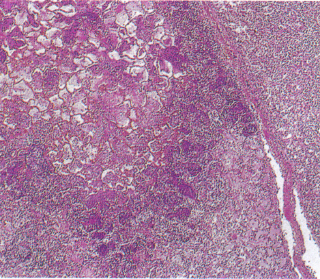
| 5) Clearly defined necrotic lesions seen in Pasteurella haemolytica (HE staining). |
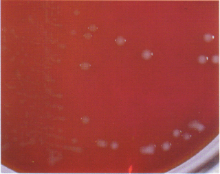
6) Colonies of P. multocida on blood agar, 37°C for 24 hrs culture. |
 |
 |
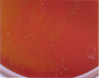
| 7) Colonies of P. haemolytica, smaller than those of P. multocida and recognizing hemolysis around the colonies (Blood agar, 37 C for 24 hrs culture). |
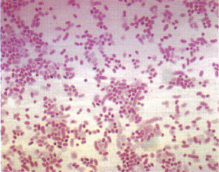
8) Bacteria of P. haemolytica (Gram staining). |
| 27 |
| -1- | -2- | -3- | -4- | -5- | -6- | -7- |